
Revolutionizing Dentistry with AI and Personalized Risk Assessment
February 27, 2023Nanomaterials in Dentistry
April 18, 2023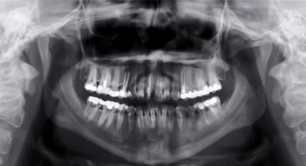
The panoramic view is a two-dimensional image that captures a panoramic view of the maxillofacial region. The image is created by rotating an X-ray source around the patient’s head, while the film or digital sensor rotates in the opposite direction. This results in a panoramic view of the entire maxillofacial region, including the teeth, jaws, sinuses, and other structures.
Panoramic radiography is particularly useful in diagnosing conditions such as impacted teeth, cysts, tumors, and fractures. It is also useful in planning orthodontic treatment and implant placement, as it provides a clear view of the underlying bone structure.
In addition to its diagnostic capabilities, the panoramic view is a valuable tool for patient education. Dentists can use the panoramic view to show patients the condition of their teeth and explain the treatment options available to them.
One of the key advantages of the panoramic view is its low radiation dose compared to other types of dental X-rays. This makes it a safer option for patients, particularly children and pregnant women.
However, the panoramic view does have some limitations. The image is two-dimensional and can result in some distortion of the image. Additionally, the panoramic view is not as detailed as other types of X-rays, such as cone beam computed tomography (CBCT).
In conclusion, the panoramic view is a valuable diagnostic tool in dentistry, providing a comprehensive view of the maxillofacial region. While it has some limitations, its low radiation dose and ability to provide a panoramic view make it an important tool in dental practice.